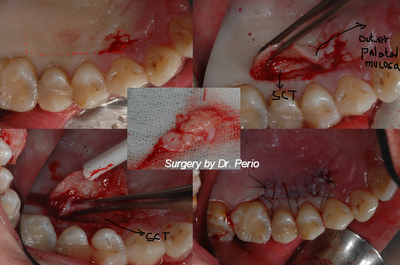
The procedure usually follows the following protocol:
1) Local anesthetic is given.
2) The tissue below the receded gum is dissected away exposing underlying tissues.
3) Gum tissue is procured from the roof of the mouth. This is usually a 1-2mm paper-thin graft that can easily be positioned around the problem tooth. The roof of the mouth is essentially an eternal reservoir of gum tissue. The donor site will be slightly tender but usually heal quickly and return to normal after a few days.
4) The graft tissue is then sewed to place over the exposed underlying tissues. Sutures are enough to secure the graft and allow for proper healing. The graft will turn first white and then often red as the graft bonds to the surrounding tissue. Ultimately, grafts will range from normal colored to slightly.
New variations of grafting have emerged over the past several years. Tissue bank skin graft material is available for those patients who would prefer not to use the roof of the mouth as a donor site. These grafts tend to have more shrinkage but will provide more natural color matching to the surrounding gums. Root coverage is now a very predictable option for teeth with receding gum lines. Subepithelial Connective Tissue Grafts borrow internal tissue from the roof of the mouth. This tissue is positioned under a flap of gum in the area of recession. The combined nutrition from the gum under and over the graft keeps the graft alive over the previously exposed root surface. The result is both an increase in the amount of protective gum tissue as well as improved esthetics. These grafts also provide the best color match with the surrounding gums.
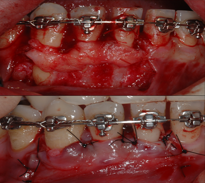
Case report: A lower anterior area generalized gingival recessions/fenestration with roots exposure during orthodontic treatment__The Subepithelial connective tissue graft to improve the thickness of overlying gingival tissue and roots coverage __Referred from other general practitioner for some malpractice during the orthodontic therapy


Periodontal diseases take on many different forms but are usually a result of a coalescence of bacterial plaque biofilm accumulation of the gingiva and teeth, combined with host immuno-inflammatory mechanisms and other risk factors which lead to destruction of the supporting bone around natural teeth. Untreated, these diseases lead to alveolar bone loss and tooth loss and, to date, continue to be the leading cause of tooth loss in adults.
ReplyDelete