


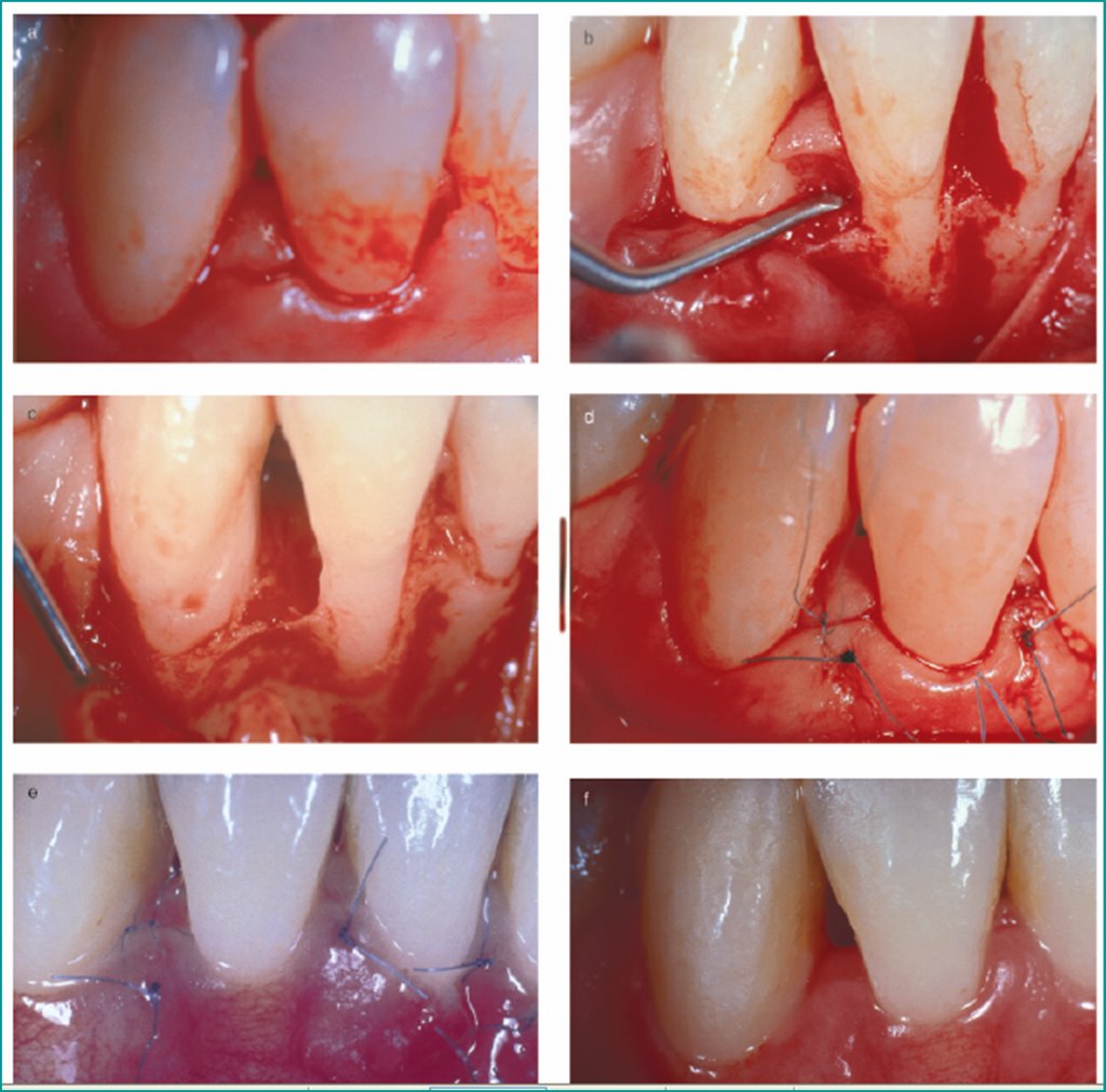
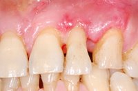
Modified Papilla Preservation Technique (MPPT)
 The modified papilla preservation technique(1996). A new surgical approach for interproximal regenerative procedures. A modification of the papilla preservation technique has been applied to achieve primary closure of the interproximal tissue over barrier membranes placed coronal
 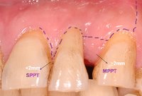 to the alveolar crest. Cortellini et al. study:Fifteen patients with deep intrabony interproximal defects were treated. Defects had a probing attachment level loss of 9.9 +/- 3.2 mm and a recession of the gingival margin of 1.7 +/- 1.6 mm. The depth of the intrabony component was 5.5 +/- 2.9 mm; while the suprabony component was 5.9 +/- 2.0 mm. Titanium-
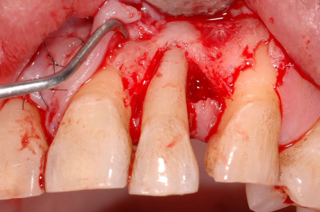 reinforced teflon membranes were placed 1.3 +/- 0.7 mm from the cemento-enamel junction, 4.5 +/- 1.6 mm coronal to the interproximal alveolar bone crest. Primary closure over the interproximal portion of the membrane was obtained in 93% of cases. In 73% of the cases complete coverage of the membrane was maintained until its removal at 6 weeks. These data indicate that the modified papilla preservation technique can be successfully applied to obtain primary closure of the interdental space in regenerative procedures with barrier membranes.
J Periodontol. 1995 Apr;66(4):261-6.
Int J Periodontics Restorative Dent. 1996 Dec;16(6):546-59.

Figure Enlarge>>>
posted by Ajou Huang @ 12:04 pm
0 comments
![]()
![]()
0 Comments:
Post a Comment
<< Home