


Maxillary Sinus Septa
Journal of Periodontology
2006, Vol. 77, No. 5, Pages 903-908
(doi:10.1902/jop.2006.050247)
 Maxillary Sinus Septa: Prevalence, Height, Location, and Morphology. A Reformatted Computed Tomography Scan Analysis
Min-Jung Kim
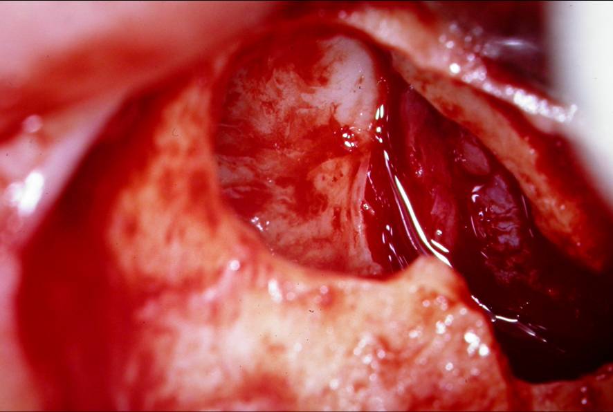
Background: The sinus lift technique may be difficult to perform if an aberrant sinus
 anatomy is encountered during surgical exposure, such as when a septum is present on the sinus floor. The objective of this study was to determine the prevalence, size, location, and morphology of maxillary sinus septa in the atrophic/edentulous and non-atrophic/dentate maxillary segments.
Methods: The sample population consisted of 100 patients (41 women and 59 men, with a mean age of 50 years, ranging between 19 and 87 years) for whom treatment was being planned for implant-supported restorations. Reformatted computerized tomograms (CT) from 200 sinuses were analyzed using imaging software.
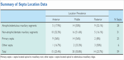
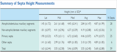
Results: The prevalence of one or more septa per sinus was found to be 26.5% (53/200), 31.76% (27/85), and 22.61% (26/115) in the overall study population and the atrophic/edentulous and the non-atrophic/dentate maxillary segments, respectively. In the analysis of the anatomic location of the septa within the sinus, it was revealed that 15 (25.4%) were located in the anterior region, 30 (50.8%) in the middle region, and 14 (23.7%) in the posterior region. The measured heights of the septa varied among the different areas. The mean heights of the septa were 1.63 ± 2.44, 3.55 ± 2.58, and 5.46 ± 3.09 mm in the lateral, middle, and medial areas, respectively.
Conclusions: It can be inferred that there is a wide anatomical variation in the prevalence, size, location, and morphology of maxillary sinus septa, irrespective of the degree of atrophy. Therefore, to prevent the likelihood of complications arising during sinus augmentation procedures, a thorough and extensive understanding of the anatomic structures inherent to the maxillary sinus is indispensable.
posted by Ajou Huang @ 9:09 pm
0 comments
![]()
![]()
0 Comments:
Post a Comment
<< Home