Click here to join perioangroup
Click to join perioangroup
Powered by health.groups.yahoo.com
Subscribe to perioangroup
Powered by health.groups.yahoo.com
Add to Google

2005-12-30

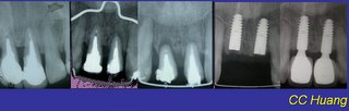
Forced Orthodontic Eruption (FOE) before Dental Implantation

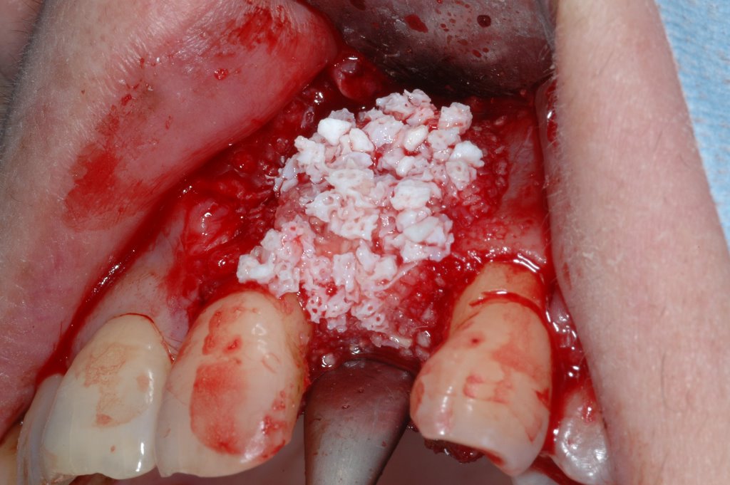
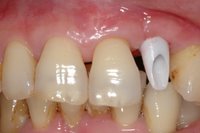
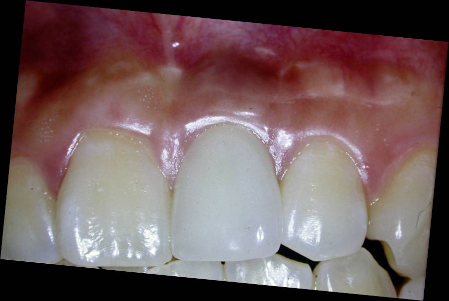
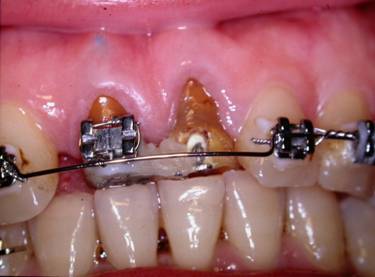
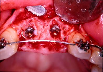
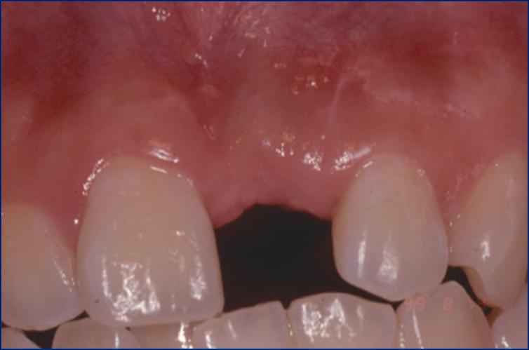
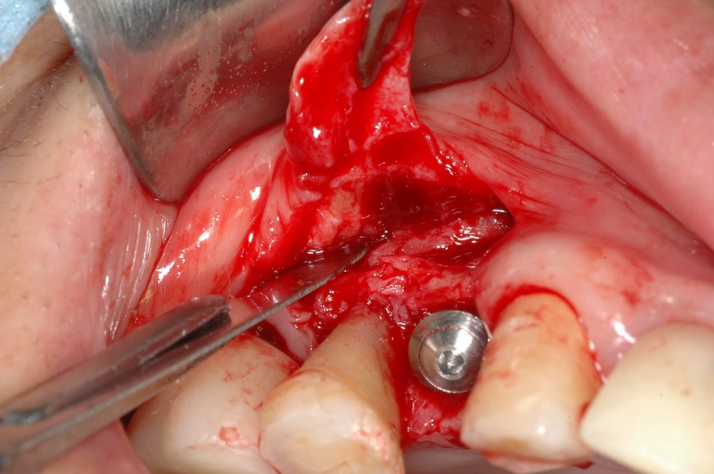
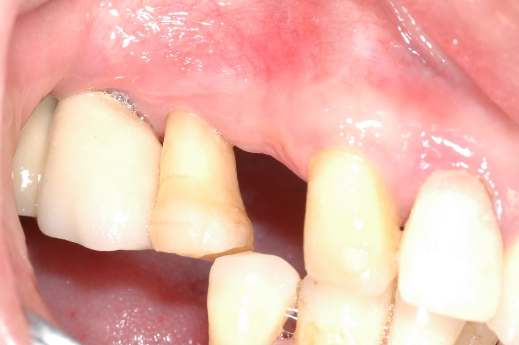
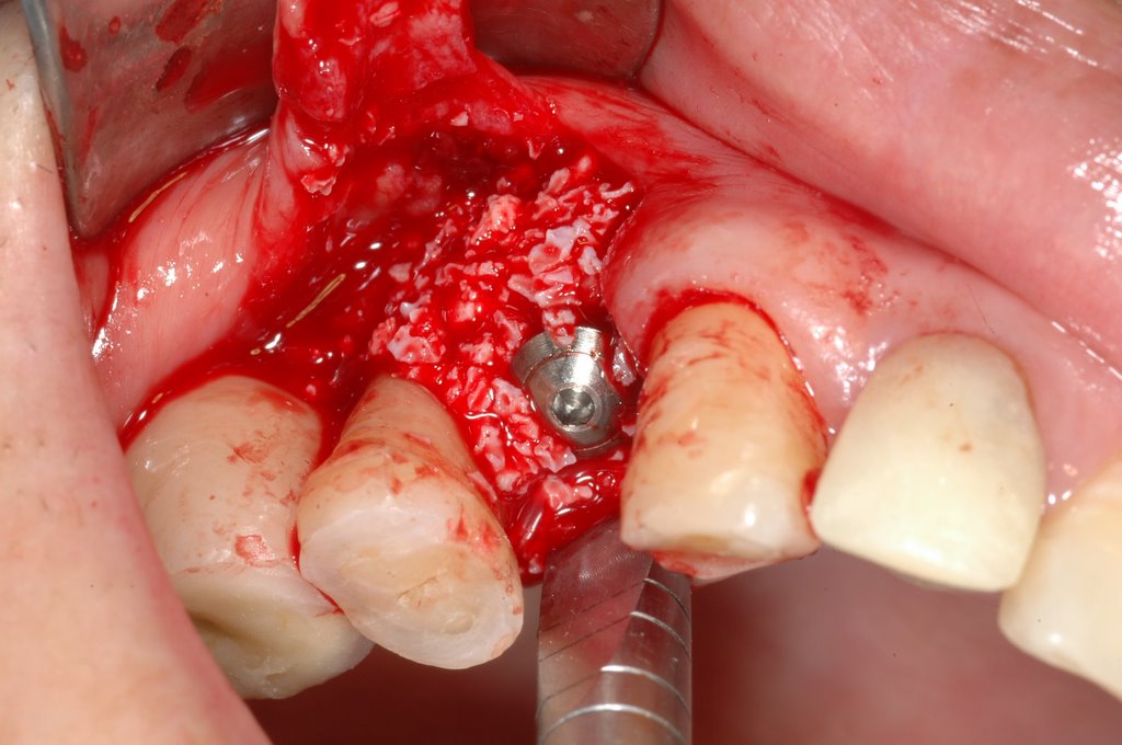
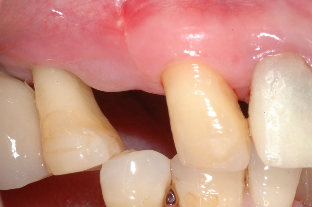
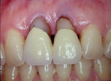
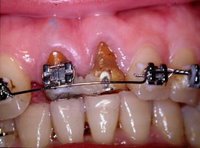
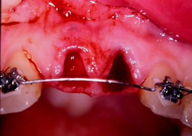
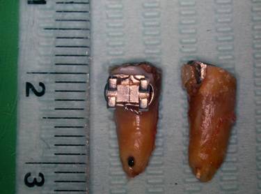
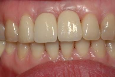
The treatment of esthetic areas with dental implants represents a new challenge for the clinician. Placementof a single-unit implant is indicated for fractured or periodontally compromised teeth. In 1993, a modification ofthe forced eruption technique, called “orthodontic extrusive remodelling,” was proposed as a way to augment both soft- and hard-tissue profiles at potential implant sites. This case report describes augmentation of the coronal softand hard tissues around the teeth with severe periodontal destruction , which was achieved by forced orthodontic extrusion before implant placement. This technique may be used to improve primary anchorage of a dental implant, fill the alveolar socket with bone, preserve interdental bone height and increase the amount of attached gingiva.
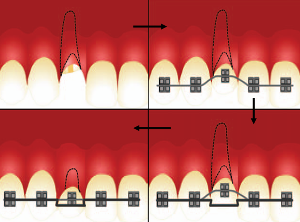
An approach to soft- and hard-tissue augmentation is forced orthodontic eruption (FOE). As first described by Heithersay and Ingber, this technique is based on osteophysiologic and orthodontic principles. It has been usedf requently because of its demonstrated advantages, including correction of isolated infrabony defects, repositioning ofthe gingival margin and clinical lengthening of thecrown.
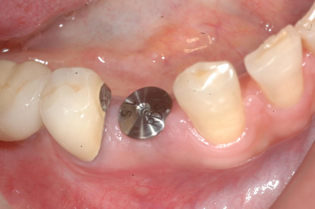
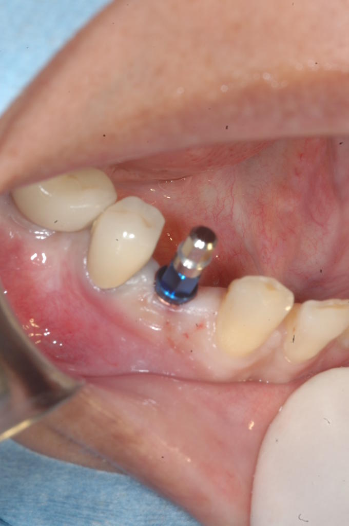
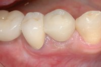
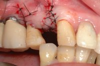
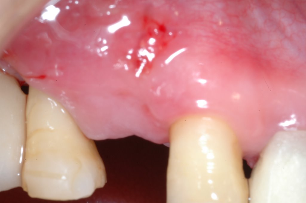
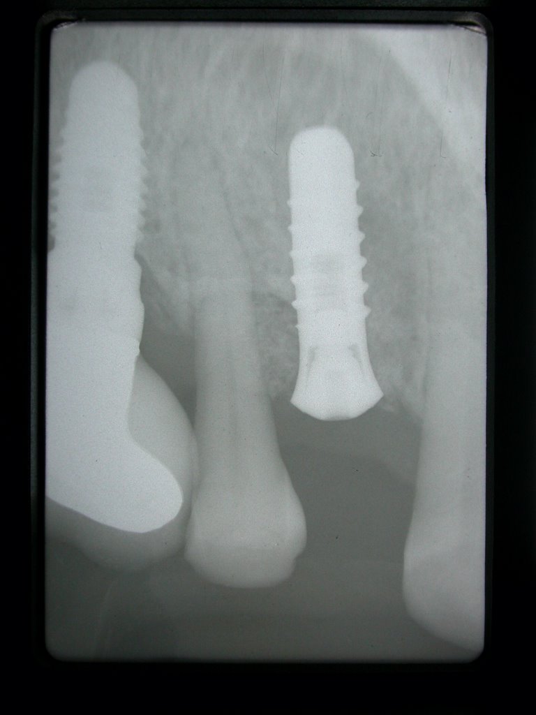
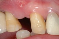
Clinical Case Report by Perio: (FOE by Dr. Tsai)


@Buccal and coronal bone augmentation using forced eruption and buccal root torque( Int J Periodontics Restorative Dent. 2003 Dec;23(6):585-91.)

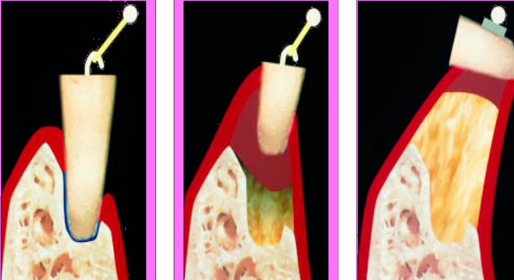
*Multidisciplinary Treatment Approach for Enhancement of Implant Esthetics(Dr. Wang 2004)

2005-12-20

Ceramic Implant Abutment - zirconium dioxide (zirconia, ZrO2)

Cercon® The quest for a biocompatible and esthetic metal free restoration, capable of multiple unit bridges and promising reliability has been the focus of research and development activity in dental materials for the last decade. The Cercon system takes advantage of the excellent strength, reliability and biocompatibility of ceramic zirconia (zirconium oxide, ZrO2), along with the accuracy, control and advance materials capability of an innovative computer aided machining (CAM) process. Cercon also allows dental professionals to bring the wealth of their clinical and lab experience to bear, to design and create esthetic metal-free restorations best suited to their patients’ need.

A case presentation with Cercon® abutment and Cercon® all-ceramic crown:(photoright by Perio)


2005-12-18

Flapless dental implantation

For esthetic outcome! Flapless approach to dental implantation is highly recommended.ATRAUMATIC EXTRACTION BY Periotome first.
Then Inserted implant into the prepared bone site.
It will keep the soft and hard tissue as much as possible. Home>>>

Dr. Buser's Opinions for Flapless appraoch of dental implantation:

2005-12-17

Anterior Implant Esthetics

The first decision to make is whether the failing tooth is needed for implant site development. The failing tooth may be useful in situations where significant bone loss is associated with the failing tooth but not with adjacent teeth. If one extracts the tooth, the bone level may be more apical relative to the adjacent teeth. This creates a difficult situation for impression taking and implant-supportive maintenance. As plaque accumulates at the implant interface, the inflammation may adversely affect the periodontal status of adjacent teeth. The ideal treatment is to delay the extraction so the failing tooth can be orthodontically extruded. As this is done over the course of six months, the bone and periodontal attachment surrounding the failing tooth will be coronally positioned to match adjacent bone levels. This provides more ideal bone height and allows the restorative implant platform to be placed at the desired height of 3 to 4 mm subgingivally. This section will focus on situations where the failing tooth needs to be extracted and define key determinants for preserving hard and soft tissue for an optimal implant-supported prosthesis.
There are several factors to consider when planning an implant restoration of failed dentition in the esthetic zone. Most often, the esthetic zone involves the maxillary anterior dentition. Although the single-tooth implant-supported restoration appears simple to perform, it is the most challenging in the esthetic zone. One of the initial assessments is determining if the tooth in question is a candidate for immediate implant replacement or a ridge preservation procedure.
The following checklist of nine issues will be discussed.
* Assessment of patient expectations;
* Assessment of gingival display;
* Gingival thickness;
* Papilla presence or absence;
* Morphology of adjacent teeth (crown-to-root ratio);
* Size and shape of contact areas of adjacent teeth;
* Available bone height;
* Available bone width; and
* Studies appropriate for final decision making.


Clinical Considerations for Increasing the Esthetic for Anterior Single-tooth Implant Restorations
1. Bone volume-quality and contour
2. Proximity of anatomical structures
3. Gingival morphology-facial and interproximal
4. Periodontal prognosis of adjacent teeth
5. Mesiodistal width of edentulous space
6. Interocclusal space
7. Implant size and site
8. Custom provisional prosthesis and abutment
9. Oral hygiene, etc.

2005-12-12

Hydraulic Sinus Condensing (HSC) for Implant Placement

The More Predictable and Less Invasive 21st Century Sinus Augmentation Technique

Background of Hydraulic Sinus Condensing (HSC)Technique

Developed by Leon Chen, D.M.D., M.S., Hydraulic Sinus Condensing, is today’s premier technique for sinus augmentation. Dr. Chen has demonstrated this technique to sold-out crowds at the annual meeting of the American Academy of Periodontology, the European Federation of Periodontology, and other academy meetings.

To read an interview about Hydraulic Sinus Condensing, please click here.

An 8-year retrospective study: 1,100 patients receiving 1,557 implants using the minimally invasive hydraulic sinus condensing technique. Chen L, Cha J.
J Periodontol. 2005 Mar;76(3):482-91

Clinical procedure:


New Technique by Dr. Smiler?? :
Balloon Subantral Membrane Elevator ( SME )
The advantages of Subantral Membrane Elevator, or SME:
Decreases Surgical Time
Reduces Risk of Membrane Tears
Mini Balloon Inflates to 3.1mm

(Micro Mini Inflates to 1.9mm)
Single Use, Supplied Sterile
Supplied with 5cc Luer Lok® Syringe
Can be used in small osteotomies


*Effects of sealing the perforated sinus membrane with a resorbable collagen membrane: a pilot study in humans.

J Oral Implantol. 2003;29(5):235-41

Proussaefs P, Lozada J, Kim J.

Graduate Program in Implant Dentistry, Loma Linda University, Loma Linda, CA 92350, USA.

The effects of repairing the perforated sinus membrane with collagen membrane are unknown. The purpose of this pilot study was to clinically, histologically, and histomorphometrically evaluate the results of repairing the perforated sinus membrane with resorbable collagen membrane. A split-mouth design was followed in the current study. Five subjects requiring bilateral sinus grafting were included in the study, where one site was accidentally perforated during sinus augmentation procedures and the other site was not perforated. The perforated sites were repaired with a resorbable collagen membrane. Dental implants were placed at a second stage and biopsies were harvested from both sinuses. New bone formation was measured for all sites. Implant survival was recorded at second-stage surgery. Nonperforated sites demonstrated significantly more bone formation (34.40%) than perforated sites (12.80%) (P = .016). Implant survival at second-stage surgery was significantly inferior in perforated sites (54.5%) when compared with nonperforated sites (100%) (P = .0146). The study demonstrated that perforation and repair of the Schneiderian membrane can compromise new bone formation and implant survival rate.

2005-12-05

Tension-Free Suture for Dental Implantation or Bone Augmentation

To obtain tension free primary closure, the buccal full thickness flap periosteum was scored using horizontal releasing incisions and spreading by Metzenbaum scissors or Scaple after insertion into the tissue. The flap was approximated using Gortex® (Gore, Flagstaff, AZ) sutures or monofilament suture. These sutures prevent wicking of bacteria into the graft as well as maintain tensile strength. One cannot overly emphasize the importance of establishing and maintaining primary closure following bone-grafting procedures. The sutures were removed two weeks postoperative. The patient agreed to refrain from wearing a prosthesis over this area thus facilitating undisturbed healing.
Clinical Technique of Tension-free Sutures by Perio:
<< post Sx 1 wk ..............postSx 2 wks>> >>PostSx 1.5mo
Join Discussion@