Click here to join perioangroup
Click to join perioangroup
Powered by health.groups.yahoo.com
Subscribe to perioangroup
Powered by health.groups.yahoo.com
Add to Google

2006-05-30

Dental Implant Fracture: a complication of treatment with dental implants

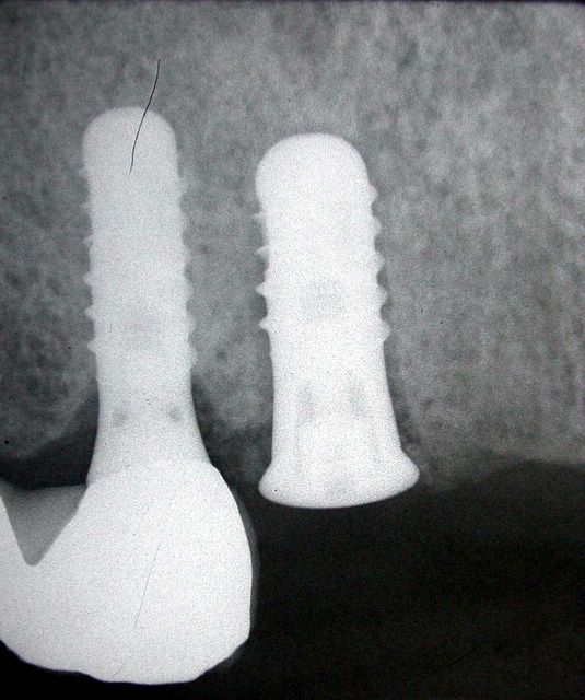
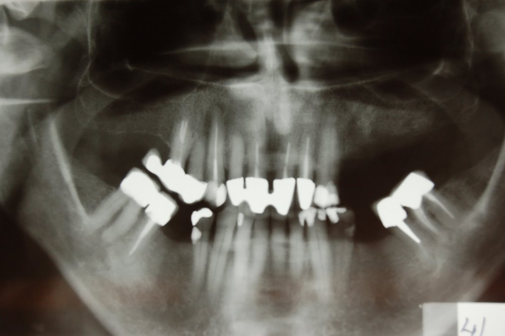
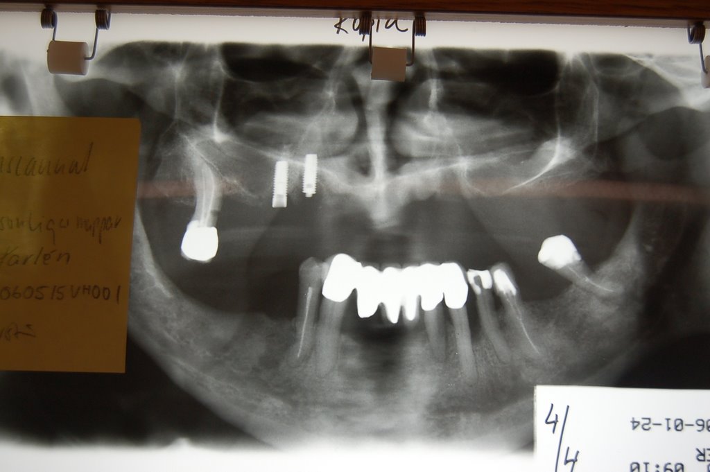
Dental implants are a functional and esthetic solution to partial and total edentulism. The initial success rate of this treatment modality is 90-95%. But, that treatment modality is not free of complications. One of the rare complications yet, with severe clinical results is fracture of dental implants. The current literature review (Biomaterials. 2002 Jun;23(12):2459-65)presents the various causative factors that may lead to implant fracture. Implant failures may be sorted into groups by the timing of their appearance, or by the origin of failure. Fractures belong to the group of late complications, caused by a biomechanical overload. 
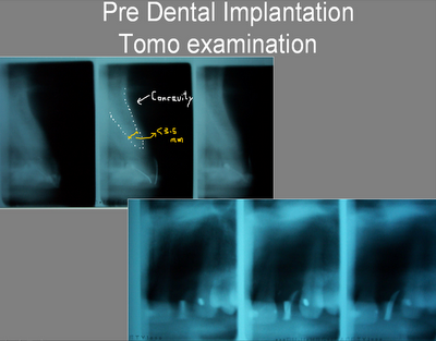
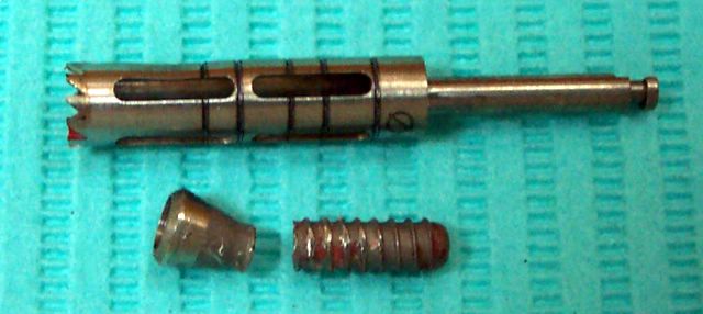
Overload may be caused by inappropriate seat of the superstructure, in-line arrangement of the implants, leverage, heavy occlusal forces (bruxing, clenching), location of the implant and the size of the implant or metal fatigue. Good clinical examinations and correct treatment plans may reduce the risk of implant fracture.
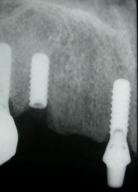
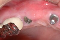
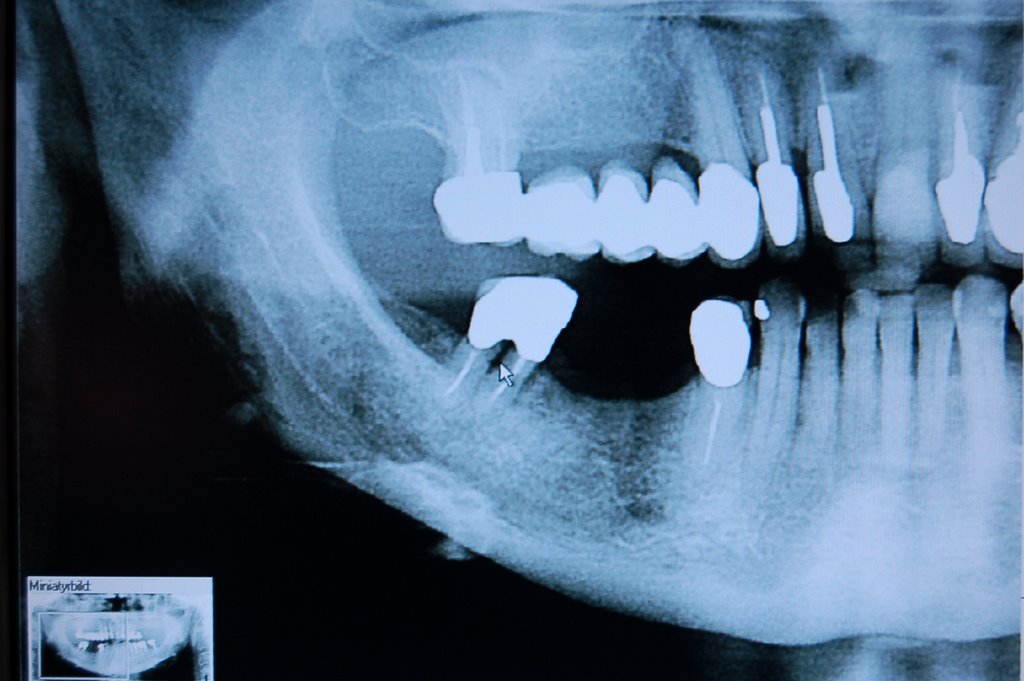
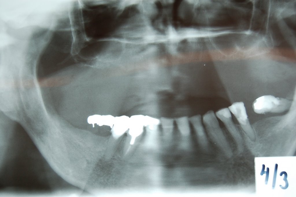
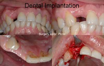
*A Case Report for Fractured Implant Removal: >>
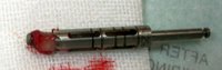
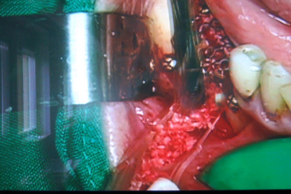
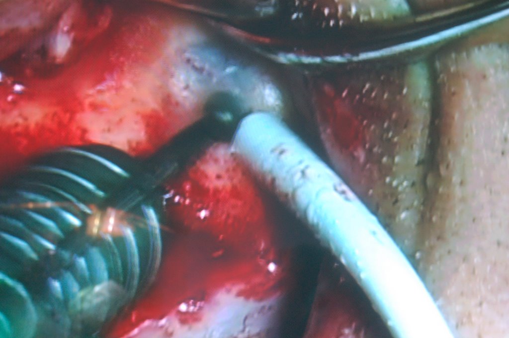
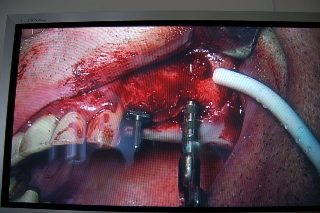
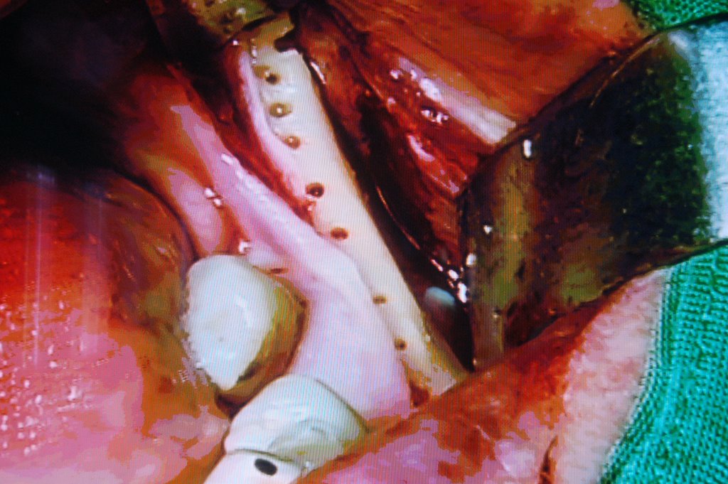
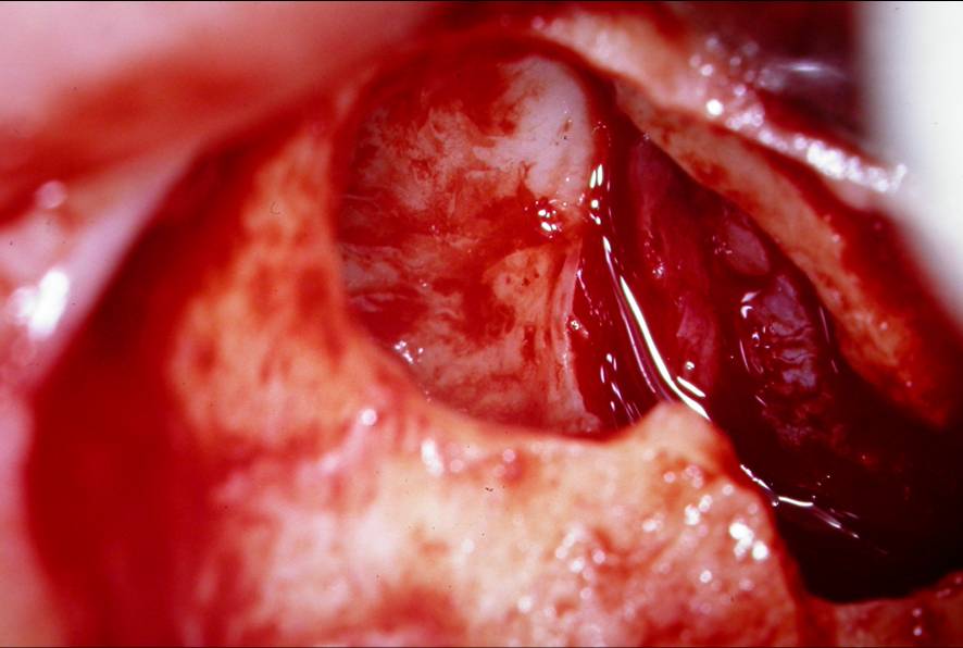
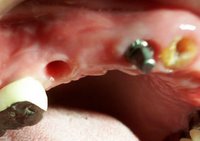
Trephine to remove the fractured implant


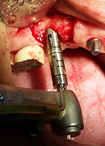
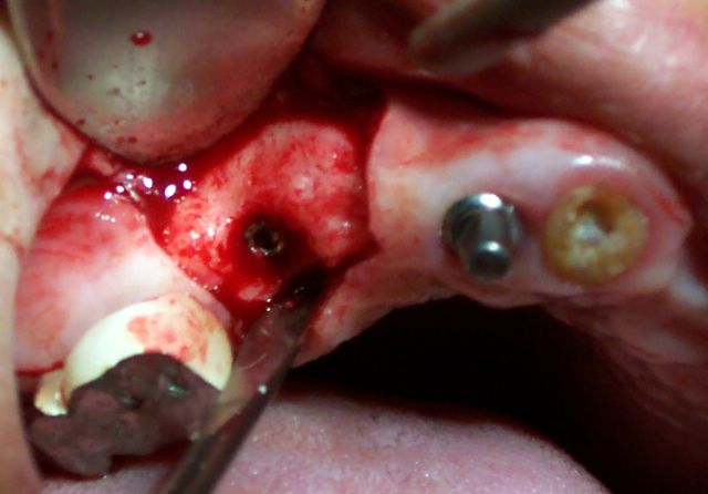
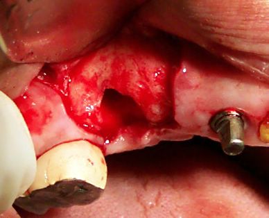
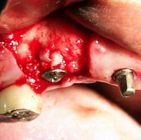
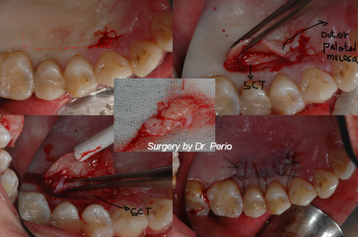
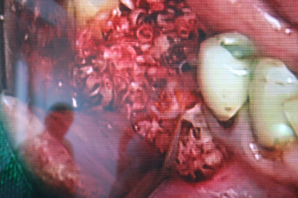
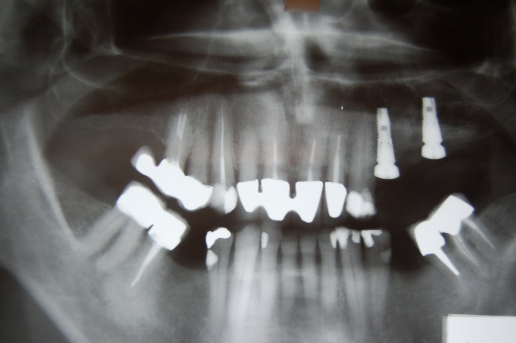
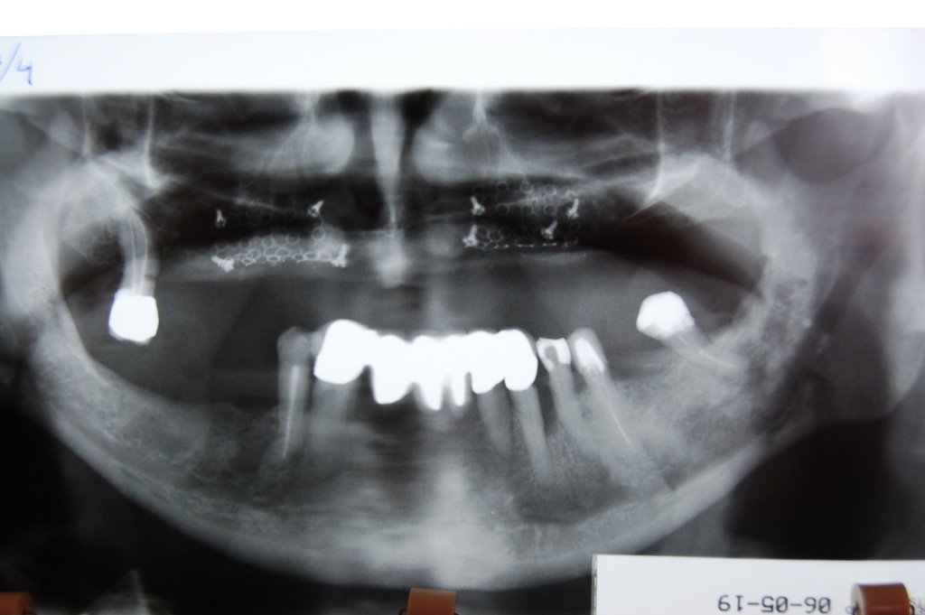
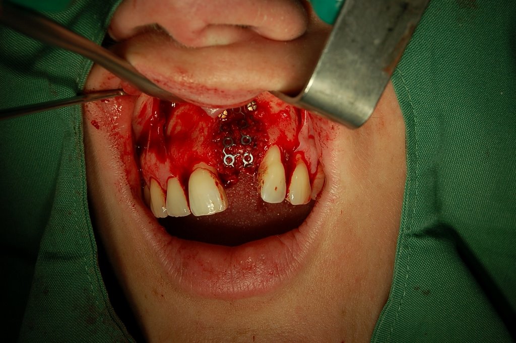
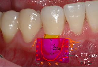
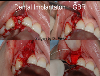
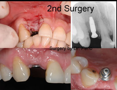
*New larger implant with bone graft into the old implant site>>


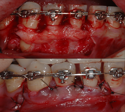
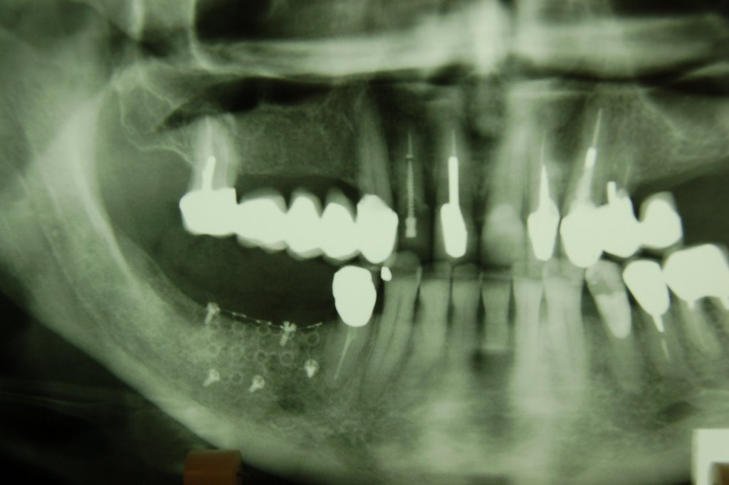
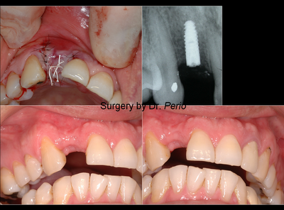
>>>one & 2.5 months later>>>

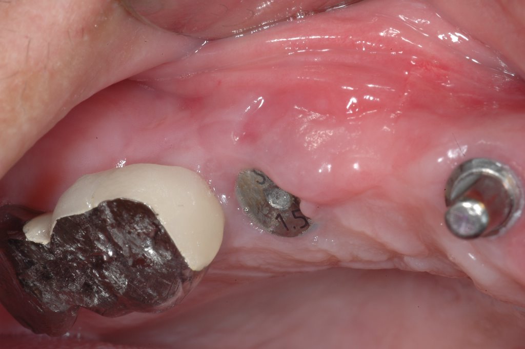
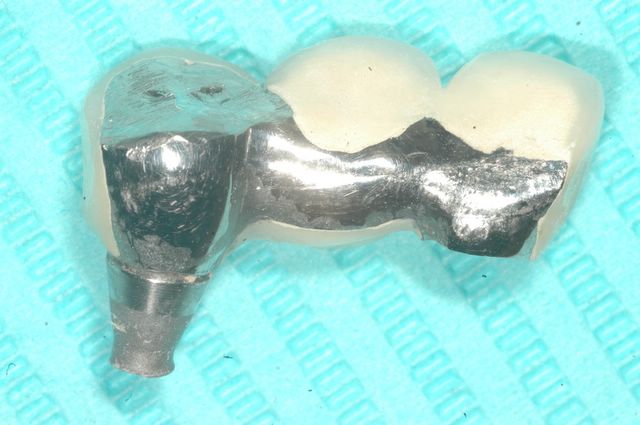
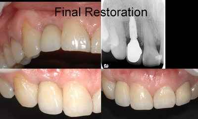
@Final Restoration:( after 6 months later from reimplantation!)
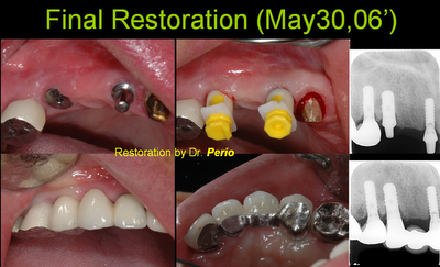
Labels:

2006-05-27

Periodontal Treatment Options for Receding Gumlines

Erosion of the gums at the gumline is relatively common. It is seen as a result of chronic inflammation associated with chronic gum disease. I t is also fairly common in young adults with aggressive toothbrushing habits. Generally, teeth will develop gum recession when a root is prominent in the bone compared to other teeth. Patients who grind their teeth also stress the sockets in which the teeth reside. When this stress is delivered to sockets with thin bone, loss of bone and gum may occur. The resulting gum recession can lead to thermal sensitivity to the tooth and may be unsightly. Dentists are concerned about recession when the amount of gum tissue deteriorates. Research has shown that when the amount of attached gum tissue diminishes below one millimeter, these teeth are more vulnerable to developing further recession. When these teeth are treated with gum grafts, the recession will stabilize and help to prevent the further loss of tooth support.Soft Tissue Grafts are procedures that rebuild the protective gum tissue around the tooth or teeth.

The procedure usually follows the following protocol:
1) Local anesthetic is given.
2) The tissue below the receded gum is dissected away exposing underlying tissues.
3) Gum tissue is procured from the roof of the mouth. This is usually a 1-2mm paper-thin graft that can easily be positioned around the problem tooth. The roof of the mouth is essentially an eternal reservoir of gum tissue. The donor site will be slightly tender but usually heal quickly and return to normal after a few days.
4) The graft tissue is then sewed to place over the exposed underlying tissues. Sutures are enough to secure the graft and allow for proper healing. The graft will turn first white and then often red as the graft bonds to the surrounding tissue. Ultimately, grafts will range from normal colored to slightly.
New variations of grafting have emerged over the past several years. Tissue bank skin graft material is available for those patients who would prefer not to use the roof of the mouth as a donor site. These grafts tend to have more shrinkage but will provide more natural color matching to the surrounding gums. Root coverage is now a very predictable option for teeth with receding gum lines. Subepithelial Connective Tissue Grafts borrow internal tissue from the roof of the mouth. This tissue is positioned under a flap of gum in the area of recession. The combined nutrition from the gum under and over the graft keeps the graft alive over the previously exposed root surface. The result is both an increase in the amount of protective gum tissue as well as improved esthetics. These grafts also provide the best color match with the surrounding gums.

Case report: A lower anterior area generalized gingival recessions/fenestration with roots exposure during orthodontic treatment__The Subepithelial connective tissue graft to improve the thickness of overlying gingival tissue and roots coverage __Referred from other general practitioner for some malpractice during the orthodontic therapy

2006-05-19

Live surgery Case 3~5

Live surgery case3:
Very narrow ridge over right mandibular -
Open type Ti mesh + Autogenous bone graft (Harvesting from Safe scaper)


@Live surgery case4: Lt' Maxillary sinus lift + Intra-antral bone grafting + Dental implantation


@Live surgery case5: Failing implant removal + Ridge augmentation / Sinus augmentation under Ti mesh


*** The Use of Ramus Grafts for Ridge Augmentation- Clinical Article-(Dental Implantology Update: June 1998)
@ Photo with professor Isaksson and Dr. Becktor

@ Ramus onlay graft harvesting procedures: Video (Please don't publish in public): http://www.youtube.com/watch?v=Xk8rstO-tME

2006-05-18

A wonderful course in a lovly city

Clinical Training Course -
Advanced implant Surgery with Forcus on Minor Bone Grafting Procedures

May 18-19, 2006
Maxillofacial Unit, Halmstad Hospital, Halmstad, Sweden

Astra Tech AB

Participants:
Dr. Ramon Gomez Meda (Spain)
Dr. Alberto Trigas Damian (Spain)
Dr. Lennart Mollersten (Sweden)
Dr. Sven-Ake Nilsson (Sweden)
Dr. Stefan Ohlsson (Sweden)
Dr. Chi-Chou Huang (Taiwan)
Dr. Yin-Pin Wang (Taiwan)

Faculty:
Sten Isaksson DDS MD PhD (Head of the maxillofacial Unit Oral & Maxillofacial Surgeon)
Jonas Becktor DDS

Two Live Surgery on First Day
#case 1
#case 2

2006-05-15

Out town 1 week to attend advanced implant surgery course in Sweden

Advanced Implant Surgery with Focus on Minor Bone Grafting Procedures

On two different occasions: May 18–19 and September 14–15, 2006 Maxillofacial Unit, Halmstad Hospital, Halmstad, Sweden. For the clinician with experience in conventional implant surgery and an interest in advanced surgical techniques.Various pre-implant surgical techniques suitable for general dentistry will be discussed and performed. The course includes local bone grafting such as sinus lifting and onlay grafting as well as distraction osteogenesis and fixture installation in grafted bone. Lectures, patient demonstrations and live surgery with opportunities for the participants to assist will illustrate the different procedures.

Lecturers:Sten Isaksson, DDS, MD, PhD, Clinical Director,Maxillofacial Unit, Halmstad Hospital, Halmstad, Sweden

Jonas Becktor, DDS, Consultant, Maxillofacial Unit, Halmstad Hospital, Halmstad, Sweden


Course ContentLectures
• Indications and limitations for local bone graftingtechniques suitable for general dentistry
• Distraction Osteogenesis (DOG)
• Treatment planning and discussions of participants’ own cases
• Postoperative treatment– Follow-up– Complications
• Various bone grafting techniques suitablefor local and general anesthetics
• Research and studies on implants and bone graftLive Surgery*
• Titanium mesh and bone graft• Bilateral sinus inlay bone graft
• Onlay bone graft for single tooth implant• Implant surgery in grafted bone

Duration: 2 days
Participants: Min 7, max 8
Course fee: SEK 11,300 ex. VAT.
Included in the fee: Course material, diploma, coffee,lunches and one dinner.
Language: English

@Welcome to my Astra nordic tour Blog: http://astratour.blogspot.com/
@ Join a special group for Astra dental implant:
http://health.groups.yahoo.com/group/astraimplant/join


Click here to join astraimplant
Click to join astraimplant

2006-05-13

How to predict the percentage of root coverage ?

@ A case (#43) of severe gum recession and root exposure after orthodontic treatment:

  1. Severe root prominence
  2. Root outer the alveolar house
  3. Miller's classification : Class III
  4. Root exposure with wide and deep type

For severe gum recession + loss of some interproximal bone + wide and deep root exposure>>> 100% root coverage is not possible!

....70~80 % root coverage will be predictale goal.

....Cervical operative restoration may be needed to cover exposed root surface.

2006-05-12

A Dentist taking the implant surgery and restoration

A handsome dentist (famous at his hometown) suffering an anterial tooth missing and flipper discomfort!
He decided to have a fixed restoration after dental impant and bone augmentation.
For his thin and bone concavity over the implant site, the bone augmentation and dental implantation should be together.
When a healing abutment into the implant at 2nd surgery, the implant position was far palatally. Lab technician (顏氏技工所) helped him to correct implant position by custom made angle abutment from bone crestal level.
Final result is OK. and he restores his shining smile again !

@Detail procedures see the following pictures:


2006-05-08

Maxillary Sinus Septa

Journal of Periodontology
2006, Vol. 77, No. 5, Pages 903-908
(doi:10.1902/jop.2006.050247)

Maxillary Sinus Septa: Prevalence, Height, Location, and Morphology. A Reformatted Computed Tomography Scan Analysis

Min-Jung Kim


Background: The sinus lift technique may be difficult to perform if an aberrant sinus anatomy is encountered during surgical exposure, such as when a septum is present on the sinus floor. The objective of this study was to determine the prevalence, size, location, and morphology of maxillary sinus septa in the atrophic/edentulous and non-atrophic/dentate maxillary segments.
Methods: The sample population consisted of 100 patients (41 women and 59 men, with a mean age of 50 years, ranging between 19 and 87 years) for whom treatment was being planned for implant-supported restorations. Reformatted computerized tomograms (CT) from 200 sinuses were analyzed using imaging software.
Results: The prevalence of one or more septa per sinus was found to be 26.5% (53/200), 31.76% (27/85), and 22.61% (26/115) in the overall study population and the atrophic/edentulous and the non-atrophic/dentate maxillary segments, respectively. In the analysis of the anatomic location of the septa within the sinus, it was revealed that 15 (25.4%) were located in the anterior region, 30 (50.8%) in the middle region, and 14 (23.7%) in the posterior region. The measured heights of the septa varied among the different areas. The mean heights of the septa were 1.63 ± 2.44, 3.55 ± 2.58, and 5.46 ± 3.09 mm in the lateral, middle, and medial areas, respectively.
Conclusions: It can be inferred that there is a wide anatomical variation in the prevalence, size, location, and morphology of maxillary sinus septa, irrespective of the degree of atrophy. Therefore, to prevent the likelihood of complications arising during sinus augmentation procedures, a thorough and extensive understanding of the anatomic structures inherent to the maxillary sinus is indispensable.