Click here to join perioangroup
Click to join perioangroup
Powered by health.groups.yahoo.com
Subscribe to perioangroup
Powered by health.groups.yahoo.com
Add to Google

2006-04-29

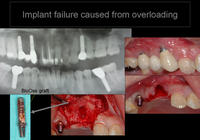
Dental Implant Overload

Dental Implant Overload
@information from the atlantadentalimplants.com
Nobody likes to be overworked. Dental implant dentists say that a dental implant is overloaded when the dental implant has too much work to do. It is always best to have one dental implant replace one missing tooth. However, dental implant patients do not always want to pay for the best treatment available and dental implant dentists try hard to please their patients. Yes, a dental implant patient can get by with less than one dental implant per missing tooth, but this is not ideal and leads to overworking the dental implants harder than the original natural teeth! In the long run, the money saved may not be worth it. In matters of health, it is always best to choose the best road, not the cheapest.
Dental implants can also be overloaded by poorly designing the dental work on top of the dental implants or by poorly placing the dental implants. Both of these problems can often be avoided with excellent dental implant treatment planning before the dental implants are ever placed. Dental implants should ideally be placed so that a dental implant patient's biting forces are directed straight downward onto the dental implant. This can not always be achieved especially when a dental implant patient has lost a great deal of bone.

@Evaluation of load transfer characteristics of five different implants in compactbone at different load levels by finite element analysis. (PDF!)

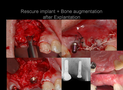
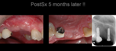
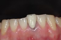
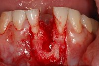
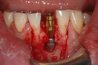
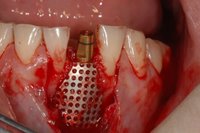
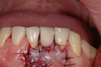
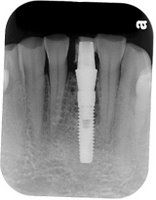
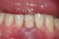
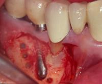
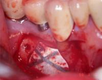
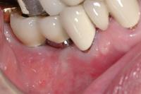
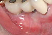
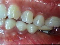
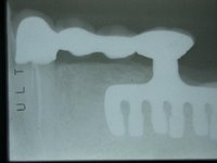
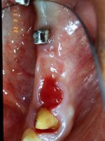
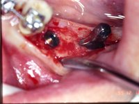
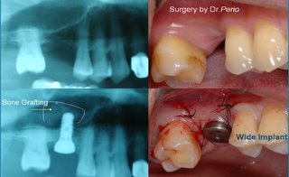
@ A case with another implant rescure and bone augmentatrion after dental implant overloading. (Surgery by Dr. Perio)

2006-04-28

Paroxysmal positional vertigo as a complication of osteotome sinus floor elevation

Eur Arch Otorhinolaryngol. 2005 Aug;262(8):631-3. Epub 2005 Feb 27.

Paroxysmal positional vertigo as a complication of osteotome sinus floor elevation.
Di Girolamo M, Napolitano B, Arullani CA, Bruno E, Di Girolamo S.
School of Dentistry, University of Rome Tor Vergata, Rome, Italy.

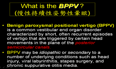
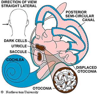
Paroxysmal positional vertigo (PPV) is a high prevalence, vestibular end organ disorder due to the detachment of the utricular otoconia floating in the posterior or lateral semicircular canal. Even though in the majority of cases the etiology of PPV is unknown, it may follow viral infection, vascular disorders and head trauma after different surgical procedures. The aim of this study was to investigate the correlation between PPV and the surgical trauma induced by the vibratory and percussive forces on the upper maxilla during the osteotome sinus floor elevation procedure. We performed a complete otoneurological examination on 146 patients affected by atrophic ridges before and after upper maxilla surgery. Four patients showed a PPV of the posterior semicircular canal controlateral to the implanted side 1 or 2 days after the surgical procedure, which promptly was solved with the Epley re-positioning maneuver. We hypothesize that the surgical trauma, and specifically the pressure exerted by the osteotomes, determines the detachment of the otoliths from the utricular macula while the patient head position, hyper-extended and tilted opposite to the side where the surgeon is working, favors the entry of these free-floating particles in the posterior semicircular canal of the implanted side. Although this disease is rather frequent in the normal population and it is a benign, self-limiting peripheral disorder, it should be considered by the oral surgeon as a possible complication of pre-prosthetic upper maxilla surgery, and the patient should be informed before undergoing surgery.

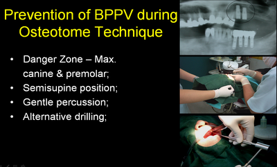
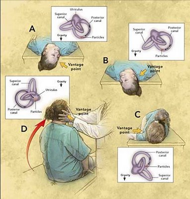
@ Clinical suggestions for preventing BPPV: (Dr. Perio had the BPPV after ridge expansion with Osteotome technique twice!) @ Dignosis and Therapy for BPPV: Epley maneuver
***Supplemental material on the site CD: Animation of Epley Maneuver.
Note that this maneuver is done faster in the animation than in the clinic. Usually one allows 30 seconds between positions

2006-04-26

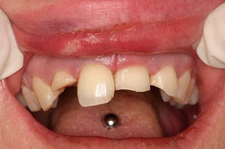
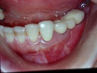
Multiple dental fractures following tongue barbell placement

** a picture download from frickr
Multiple dental fractures following tongue barbell placement: a case report

The number of adolescents and young adults undergoing intra-oral piercing, is increasing worldwide. There have been several case reports documenting oral and systemic complications of this practice. These include damage to the dentition, gingivae, infection, speech impediments and nerve damage. The case presented here draws attention to the possibility of multiple tooth fracture as a result of trauma incurred from a barbell inserted into the tongue.

## Worldwide warning for Tongue Piercing:
@A complication of tongue piercing. A case reportand review of the literature (PDF)
@Investigating tongue piercing (BDJ)
@Tongue piercing: Case report and review of currentp r a c t i c e (ADA in Australia)
@Tongue Piercing andAssociated ToothFracture (cda-adc.CA)
@Oral piercing and oral trauma in a New Zealand sample

2006-04-25

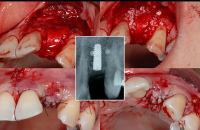
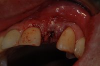
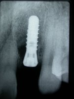
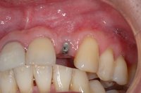
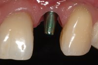
Stimutainous dental implantation with other techniques

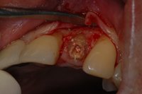
  • Presurgery - Long-term apical lesion with persistant sinus teact

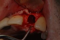
  • Atrumatic extraction and socket curettage
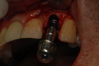
  • Immediate dental implantation
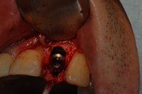
  • GBR for periimplant bony defect with BioOss bone graft + Ti mesh + Teruplug
  • Immediate provionalization

  • Post Sx 2 wks

2006-04-23

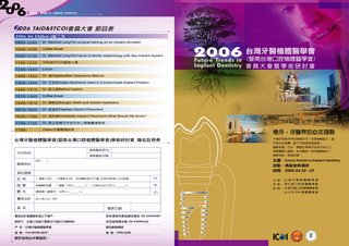
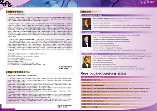
台灣牙醫植體醫學會暨南台灣口腔植體醫學會2006年年會

台灣牙醫植體醫學會暨南台灣口腔植體醫學會2006年年會

將於高雄金典酒店舉辦
時間:2006 4/22~23(六,日),
地點:高雄金典酒店 41樓
主辦:台灣牙醫植體醫學會
承辦:南台灣口腔植體醫學會
協辦:中華民國口腔植體醫學會
   台北市牙科植體醫學會
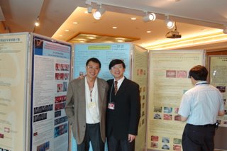
@ Dr. Perio present a post at academy meeting (Apr22~23,06)

2006-04-21

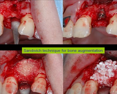
"Sandwich" bone augmentation technique

Int J Periodontics Restorative Dent. 2004 Jun;24(3):232-45.

"Sandwich" bone augmentation technique: rationale and report of pilot cases.
Wang HL, Misch C, Neiva RF.

The aim of this article is to present a new technique for augmentation of deficient alveolar ridges and/or correction of osseous defects around dental implants. Current knowledge regarding bone augmentation for treatment of osseous defects prior to and in combination with dental implant placement is critically appraised. The "sandwich" bone augmentation technique is demonstrated step by step. Five pilot cases with implant dehiscence defects averaging 10.5 mm were treated with the technique. At 6 months, the sites were uncovered, and complete defect fill was noted in all cases. Results from this pilot case study indicated that the sandwich bone augmentation technique appears to enhance the outcomes of bone augmentation by using the positive properties of each applied material (autograft, DFDBA, hydroxyapatite, and collagen membrane). Future clinical trials for comparison of this approach with other bone augmentation techniques and histologic evaluation of the outcomes are needed to validate these findings.

Mucogingival pouch flap for sandwich bone augmentation: technique and rationale.

Implant Dent. 2005 Dec;14(4):349-54
Park SH, Wang HL

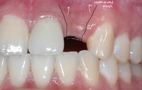
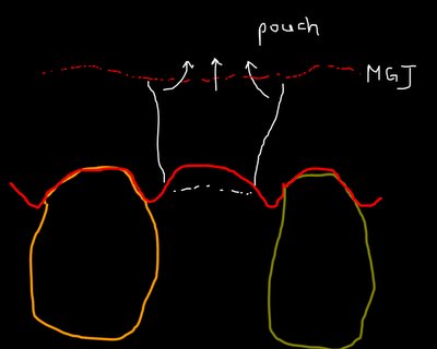
This article introduces a novel flap design, mucogingival pouch flap (MPF), to enhance the clinical outcome of sandwich bone augmentation. MPF uses a pouch flap reflection via mucogingival junction extension incisions to provide an improved graft retention, minimized membrane exposure, preserved papilla dimension, and soft tissue camouflage for improved esthetics.There are 4 implant-associated buccal dehiscence defects in 3 patients treated with sandwich bone augmentation technique in conjunction with MPF. All cases yielded an adequate new bone thickness of 1.5-3.5 mm as well as a height of 84% to 100% at 6 months. Rationales, indications, contraindications, advantages, and disadvantages for MPF designs are further discussed.

2006-04-20

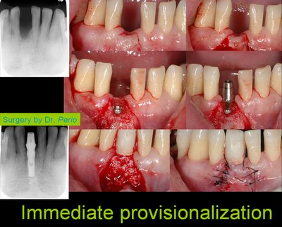
Immediate provisionalization after dental implantation

@ Single tooth immediate provisional restoration of dental implants: technique and early results.
J Oral Maxillofac Surg. 2004 Sep;62(9):1131-8
Block M, Finger I, Castellon P, Lirettle D

PURPOSE: Patients desire efficient restoration of missing teeth. Immediate provisionalization of implants at the time of placement can provide the patient with a tooth-like restoration. Our hypothesis is that preoperative fabrication of the implant abutment and provisional restoration can provide successful immediate provisionalization of implants, if specific diagnostic criteria are used for patient selection.
PATIENTS AND METHODS: This hypothesis is evaluated by prospectively following 74 implants thus treated for 6 months to 2 years. A technique is presented to illustrate a simple and reliable method to provisionally restore a single tooth restoration. The method involves preoperative placement of an implant analog into a model, preparation of the abutment on the model, and fabrication of a provisional crown out of occlusion. At the time of surgery, the implant is placed according to the prescription of the restorative dentist, the surgeon places the abutment and provisional crown, and the final restoration is fabricated after the implant integrates.
RESULTS: Seventy of 74 (94.6%) restorations have been successful with up to 2-year follow-up, which is similar to single tooth implants treated using a 2-stage protocol.
CONCLUSIONS: Single tooth immediate provisionalization implants are effective techniques when specific diagnostic criteria are used.

@Single-tooth replacement in the aesthetic zone with immediate provisionalization: fourteen consecutive case reports.

2006-04-19

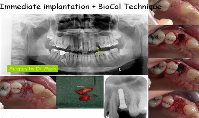
Immediate dental implantation + Bio-Col socket augmentation

Immediate Implantation in Fresh Extraction Sockets. A Controlled Clinical and Histological Study in Man

Dr. Michele Paolantonio et al.
Journal of Periodontology
2001, Vol. 72, No. 11, Pages 1560-1571

Background: Early implantation may preserve the alveolar anatomy, and the placement of a fixture in a fresh extraction socket helps to maintain the bony crest. Although a number of clinical studies exist, no histological reports show the outcome of implantation in fresh extraction sockets without the use of membranes in humans compared to implants placed in mature bone.

Methods: Forty-eight healthy patients, receiving at least 4 fixtures in each of 2 symmetrical quadrants, underwent placement of 1 experimental fixture placed in a fresh extraction socket (TI) and 1 contralateral fixture in mature bone (CI). TI were placed after atraumatical tooth extraction, with a surgical site at the apex of the socket and a tight contact between the fixture and the socket's walls, but without the use of filling materials or membranes. The flap was coronally repositioned to obtain primary wound closure. Immediately after surgical intervention, a standardized periapical radiograph was taken. Second-stage surgery was done after 6 months. Six months after the second surgery, a second standardized periapical radiograph was taken and clinical parameters (bleeding and plaque index) recorded. Marginal bone loss (MBL) from the time of implant placement to the time of fixture removal was calculated by comparing periapical radiographs. TI and CI were then removed by a hollow drill to obtain histological specimens. Non-demineralized sections were stained by acid fuchsin and toluidine blue, and by von Kossa to evaluate the degree of bone mineralization. The percentage of direct implant-bone contact (DBC) was calculated by a computerized microscopic digitizer.

Results: No significant differences in the clinical and radiographic parameters were observed between the 2 experimental categories. There was no statistically significant difference between TI and CI for DBC either in the maxilla or in the mandible. No connective or fibrous tissues were present around TI or CI. Bone resorption was not present in any of the histological sections.

Conclusions: The present study shows that when a screw-type dental implant is placed without the use of barrier membranes or other regenerative materials into a fresh extraction socket with a bone-to-implant gap of 2 mm or less, the clinical outcome and degree of osteointegration does not differ from implants placed in healed, mature bone. J Periodontol 2001;72:1560-1571

@ Clinical procedure for Immediate dental implantation after premolar tooth extraction + Socket augmentation with BioOss bone graft /Collage plug Bio-Col technique

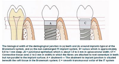
Have the " biological width " around the dental implants?

*An osseointegrated implant restoration may closely resemble a natural tooth.However, the absence of a periodontal ligament and connective tissue attachment via cementum, results in fundamental differences in the adaptation of the implant to occlusal forces, and the structure of the gingival cuff. @ The Outlines from Keynote speech by Dr. Berglundh on EAO05'
Topic:

What determines the biological width at implants?
Integrated esthetics – a biological and biomechanical approach

• Definition of “Biological width”
• Clinical soft tissue dimensions
• True soft tissue dimensions
• Soft tissue integration to different materials

Dimensions and relations of the dentoginigval junction in humans (1)
Sample:
Human autopsy specimens (Orban's and Kronfei's collections) exhibiting varying degree of periodontal tissue breakdown
Examination:
Histometric assessment of
Epithelial attachment
Connective tissue attachment

Dimensions and relations of the dentoginigval junction in humans (2)
Normal
Moderate breakdown
Advanced breakdown

Dimensions and relations of the dentoginigval junction in humans (3)
Results;
Epithelial attachment varied:
1.4 mm (normal)
0.8 mm (moderate breakdown)
0.7 mm (advanced breakdown)
CT attachment was stable:
1.1 mm
And did not vary with degree of bone loss

Dimensions and relations of the dentoginigval junction in humans (4)
Conclusion:
The biological width of soft tissue attachment is about 2.5 mm and includes 1.5 mm epithelium and 1 mm connective tissue.

Clinical soft tissue dimensions

Implant supported single tooth replacements compared to contra-lateral natural teeth.
Probing >>Chang et al 1999
Dimensions of peri-implant mucosa at evaluation of maxillary anterior single implant in man.
Bone sounding>>Kan 2003

The height of the mucosa at single implants and teeth
Implant Teeth
facial aspect
Probing: 3 mm 2 mm
Sounding: 4 mm 3 mm
proximal aspects
Probing: 4 mm 3 mm
Sounding: 5-6 mm 4 mm

The biological width: 2.5 mm (Gargiulo et al 1961)
PPD under estimates
Sounding over estimates
“Biological width” is not synonymous with Probing Pocket Depth or with Sounding

True soft tissue dimensions

The soft tissue barrier at implant and teeth (Berglundh et al 1991)

PM/GM~aJE= 2.14 / 2.05 mm
aJE~B= 1.66 / 1.12 mm
The mucosal at implants is comprised of a 2 mm long barrier epithelium and a 1-1.5 mm zone of “connective tissue integration”.
The “connective tissue integration” zone is characterized by absence of blood vessels and a large number of fibroblasts interposed between thin collagen fibers.

Dimensions of the periimplant mucosa (B & L 1996)
3 beagles dogs
3 months:
Flap adaptation and suturing
Test OE=2 mm
Control OE=4 mm
6 months:
Test
PM~aJE 2.0
aJE~B 1.3
Control
PM~aJE 2.1
aJE~B 1.8
A minimum width of the periimplant mucosa is required and bone resorption may take place to allow a proper mucosal attachment to form.

Morphogenesis of the mucosal attachment at implants (Berglundh et al 2006)
The soft tissue dimension – the biological width – at implant was established after 6 wks following Sx

How to preserve the marginal bone and avoid soft tissue recession ?
Respect the biological wwidth

Soft tissue integration to different abutment materials (Wennstom 2006)
Abutment shift (1 month after implant surgery)
Control abutment: Ti (c.p. Ti)
Test abutment: Direct (c.p. Ti)
Conclusions
The soft tissue dimensions were similar at implant abutment made of c.p. Titantium, ZrO2, based Ceramic and Au/Pt alloy
The connective tissue interface at Au/Pt alloy abutment contained lower amount of collagen and larger portions of leukocytes than that at abutment made of Ti and ZrO2

@ A review article of "teeth and implant" on BJD
@The effect of subcrestal placement of the polished surface of implants on marginal soft and hard tissues-a retrospective clinical study
*** On Line Internet Course: Biological Width by Dr. S. Robert Davidoff
@ Abstracts of Biological Width for Dental Implants
$$$ The biological aspects of the soft tissue – Titanium implant interface (PDF)
2006-04-17

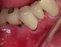
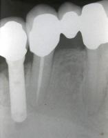
Retrograde peri-implantitis treatment

The term retrograde peri-implantitis has just recently been introduced through several case reports. It is defined as a clinically symptomatic peri-apical lesion (diagnosed as a radiolucency) that develops shortly after implant insertion while the coronal portion of the implant achieves a normal bone to implant interface (for a review, see Quirynen et al. 2003). A retrograde peri-implantitis is often accompanied by symptoms of pain, tenderness, swelling, and/or the presence of a fistulous tract . It should be distinguished from a clinically asymptomatic, peri-apical radiolucency, which is usually caused by placing implants that are shorter than the drilled cavity or by a heat-induced aseptic bone necrosis. Retrograde peri-implantitis can result from bacterial contamination during insertion, premature loading leading to bone micro-fractures, or the presence of a pre-existing inflammation (bacteria, inflammatory cells, and/or remaining cells from a cyst, granuloma). A peri-apical lesion from a nearby devitalized tooth, on the other hand, can encroach upon the implant and contaminate it (e.g. reactivation of a dormant peri-apical lesion or removal of the peri-apical endodontic seal). The treatment of periapical peri-implantitis is still empiric. The longitudinal study, together with the outcome in some case reports, seem to indicate that the removal of all granulation tissue is sufficient to arrest the progression of the bone destruction. The removal of the apical part of the implant does not seem mandatory.


Clinically stable after 9.5 years!>>

2006-04-14

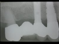
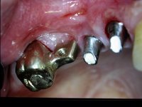
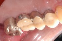
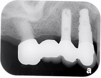
Can Dental Implants Connect with Natural Teeth?

The literature in the peer-reviewed journals seems divided on the question of connecting dental implants to natural teeth in fixed partial dentures. The problem encountered is the submersion of natural teeth producing a gap between the fixed partial denture crown margin and the prepared tooth. Some literature supports the position that dental implants and natural teeth should not be connected. Some literature supports the position that dental implants and natural teeth can be connected with rigid attachments (solid metal framework, solder joint) but not with non-rigid attachments (precision attachments).
>>>Rigid attachment with T-block>>

@Osseonews discussion for implant to natural tooth connecting

Implant to Natural Tooth Splinting
Doctors who use this philosophy will sometimes connect an implant to a natural tooth. The advantage of this is that by connecting implants to natural teeth, fewer implants are needed to complete the case. This can dramatically reduce the cost of treatment while allowing the patient to have permanent teeth. The disadvantage of this type of treatment is that should a problem arise with either the implant or natural tooth the problem has to be handled differently because the implants and natural teeth are connected. Furthermore, there are limited data regarding the effects of splinting implants to natural teeth. In this regard, it has been reported that intrusion of splinted teeth and pronounced vertical bone loss around implant abutments are potential sequelae;however, the majority of patients, , in one study suffered no adverse effects. Other reports have indicated that connecting implants to teeth in a fixed prosthesis has a good prognosis. A 5-year prospective study designed to compare bridges supported only by implants with bridges supported by both implants and natural teeth within the same patient, noted no higher risk of implant or prosthetic failure for tooth-implant fixed bridges as comparedwith implant-supported bridges.

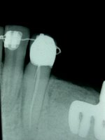
@Post Implant-Tooth connection 6 years >>>>>

OsseoNews discussion about Implant - Tooth connection


>>> Somebody did the case with implant connecting natural tooth, let the canine intrusion.<<<

>>>Orthodontic extrusion/Crown lengthening procesure of #33 + Additional implant of #34>>>

Dental implants placement in conjunction with osteotome sinus floor elevation


Dental implants placement in conjunction with osteotome sinus floor elevation: a 12-year life-table analysis from a prospective study on 588 ITI®implants
Authors: Ferrigno, Nicola1; Laureti, Mauro1; Fanali, Stefano1
Source: Clinical Oral Implants Research, Volume 17, Number 2, April 2006, pp. 194-205(12)
Publisher:Blackwell Publishing

The purpose of this prospective study was to evaluate the clinical success of placing ITI dental implants in the posterior maxilla using the osteotome technique. Material and methods:
All implants were placed following a one-stage protocol (elevating the sinus floor and placing the implant at the same time). Five hundred and eighty-eight implants were placed in 323 consecutive patients with a residual vertical height of bone under the sinus ranging from 6 to 9 mm. The mean observation follow-up period was 59.7 months (with a range of 12–144 months). This prospective study not only calculated the 12-year cumulative survival and success rates for 588 implants by life-table analysis but also the cumulative success rates for implant subgroups divided per implant length and the percentage of sinus membrane perforation were evaluated. Results:
The 12-year cumulative survival and success rates were 94.8% and 90.8%, respectively. The analysis of implant subgroups showed slightly more favourable cumulative success rates for 12 mm long implants (93.4%) compared with 10 and 8 mm long implants (90.5% and 88.9%, respectively). During the study period, only 13 perforations of the Schneiderian membrane were detected with a perforation rate of 2.2% (13 perforations/601 treated sites). Ten perforations out of 13 were caused during the first half of the study period and of these, seven were detected during the first 3 years of this prospective study.
Conclusion:
Based on the results and within the limits of the present study, it can be concluded that ITI implant placement in conjunction with osteotome sinus floor elevation represents a safe modality of treating the posterior maxilla in areas with reduced bone height subjacent to the sinus as survival and success rates were maintained above 90% for a mean observation period of ≈60 months. Shorter implants (8mm implants) did not significantly fail more than longer ones (10 and 12 mm implants): the differences were small compared with the number of events; hence, no statistical conclusion could be drawn. But, from the clinical point of view, the predictable use of short implants in conjunction with osteotome sinus floor elevation may reduce the indication for complex invasive procedures like sinus lift and bone grafting procedures .

2006-04-04

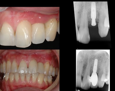
A tooth can tell a long story

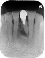
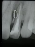
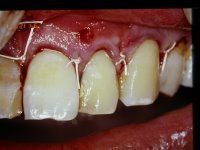
1. GTR after endodontic failure with DFDBA + Gore-Tex nonresorbable membrane
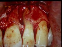
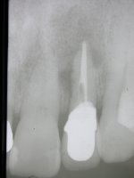
2. Root fracture after crown insertion (7 years)

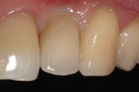
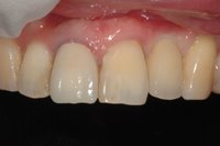
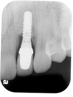
3. Immediate dental implantation after root extraction


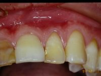
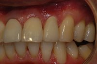
4. Final restoration